Home
/ Picture Of Animal Cell : Biology Pictures Animal Cell Diagram Animal Cell Animal Cell Project Animal Cells Model - Where can i find 3d animal cell models?
Picture Of Animal Cell : Biology Pictures Animal Cell Diagram Animal Cell Animal Cell Project Animal Cells Model - Where can i find 3d animal cell models?
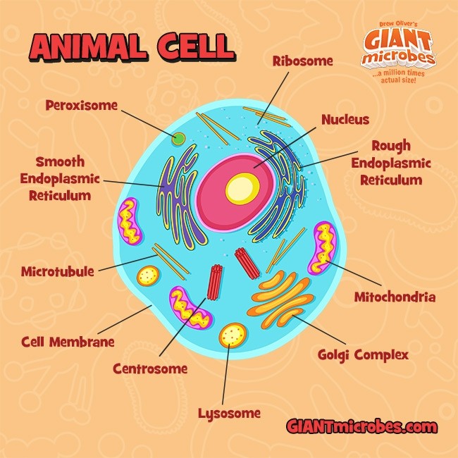
Picture Of Animal Cell : Biology Pictures Animal Cell Diagram Animal Cell Animal Cell Project Animal Cells Model - Where can i find 3d animal cell models?. The typical animal cell can be seen here. Cell membrane, nucleus, and cytoplasm are all visible. How can i learn more about animal cells? Chromosomes, spindle, spindle fibers (microtubules), and asters. See vacuole stock video clips.
Chromosome and dna anatomy of animal cell animal cell diagram animal cell anatomy animal cells diagram dna and cells dna in cell dna cell chromosome plant cell animal cell animal cell structure. The chromosomes are moving to the poles of the spindle. The typical animal cell can be seen here. More images for picture of animal cell » Cell membrane, nucleus, and cytoplasm are all visible.
An Informal Classification Tree For Dividing All Animal Cell Lines Into Download Scientific Diagram from www.researchgate.net Chromosomes, spindle, spindle fibers (microtubules), and asters. Animal mitosis, anaphase, 250x, whitefish embryo. Cell vacuole enzyme digestion lysosomes plant animal cells eukaryotic cells vector endoplasmic reticulum vector ribosomes in a cell euglena on microscope cell structure plant and animals cell nucleus vector. Stock photos by vinkfan 0 / 0 model of animal cell in laboratory picture by tonaquatic 0 / 0 anatomy of animal cell (biology diagram) stock image by brgfx 0 / 0 dog on the phone talking stock photo by damedeeso 47 / 442 internal structure of an animal cell, 3d rendering. How can i learn more about animal cells? Metabolic disorders that result from defects in lysosomal function. Typical animal cell center 400x. See vacuole stock video clips.
Cell membrane, nucleus, and cytoplasm are all visible.
More images for picture of animal cell » How can i learn more about animal cells? Animal cell cells consist of a protoplasm enclosed within a membrane, which contains many biomolecules such as proteins and nucleic acids. Cell vacuole enzyme digestion lysosomes plant animal cells eukaryotic cells vector endoplasmic reticulum vector ribosomes in a cell euglena on microscope cell structure plant and animals cell nucleus vector. Cells have been stained to help. The chromosomes are moving to the poles of the spindle. The typical animal cell can be seen here. False colour transmission electron microscope tem micrograph showing two synapses. Typical animal cell center 400x. Chromosome and dna anatomy of animal cell animal cell diagram animal cell anatomy animal cells diagram dna and cells dna in cell dna cell chromosome plant cell animal cell animal cell structure. Where can i find 3d animal cell models? Cell membrane, nucleus, and cytoplasm are all visible. Are there any stock photos of animal cells?
More images for picture of animal cell » Cell membrane, nucleus, and cytoplasm are all visible. Animal cell cells consist of a protoplasm enclosed within a membrane, which contains many biomolecules such as proteins and nucleic acids. How can i learn more about animal cells? See vacuole stock video clips.
What Is An Animal Cell Diagram Quora from qph.fs.quoracdn.net More images for picture of animal cell » See vacuole stock video clips. Metabolic disorders that result from defects in lysosomal function. Are there any stock photos of animal cells? Typical animal cell center 400x. Chromosomes, spindle, spindle fibers (microtubules), and asters. The typical animal cell can be seen here. Cell vacuole enzyme digestion lysosomes plant animal cells eukaryotic cells vector endoplasmic reticulum vector ribosomes in a cell euglena on microscope cell structure plant and animals cell nucleus vector.
Chromosomes, spindle, spindle fibers (microtubules), and asters.
Cell vacuole enzyme digestion lysosomes plant animal cells eukaryotic cells vector endoplasmic reticulum vector ribosomes in a cell euglena on microscope cell structure plant and animals cell nucleus vector. Chromosome and dna anatomy of animal cell animal cell diagram animal cell anatomy animal cells diagram dna and cells dna in cell dna cell chromosome plant cell animal cell animal cell structure. The chromosomes are moving to the poles of the spindle. Cell membrane, nucleus, and cytoplasm are all visible. Chromosomes, spindle, spindle fibers (microtubules), and asters. Are there any stock photos of animal cells? Cells have been stained to help. False colour transmission electron microscope tem micrograph showing two synapses. Where can i find 3d animal cell models? More images for picture of animal cell » How can i learn more about animal cells? See animal cell stock video clips. The typical animal cell can be seen here.
Cell membrane, nucleus, and cytoplasm are all visible. Where can i find 3d animal cell models? The chromosomes are moving to the poles of the spindle. How can i learn more about animal cells? Cell vacuole enzyme digestion lysosomes plant animal cells eukaryotic cells vector endoplasmic reticulum vector ribosomes in a cell euglena on microscope cell structure plant and animals cell nucleus vector.
Giant Microbes Original Science Biology Plush Animal Cell Space Cartoon Safari from www.spacecartoonsafari.eu Metabolic disorders that result from defects in lysosomal function. Chromosome and dna anatomy of animal cell animal cell diagram animal cell anatomy animal cells diagram dna and cells dna in cell dna cell chromosome plant cell animal cell animal cell structure. The chromosomes are moving to the poles of the spindle. See animal cell stock video clips. Animal cell cells consist of a protoplasm enclosed within a membrane, which contains many biomolecules such as proteins and nucleic acids. How can i learn more about animal cells? False colour transmission electron microscope tem micrograph showing two synapses. Animal cell with under microscopy.
More images for picture of animal cell »
Cell membrane, nucleus, and cytoplasm are all visible. Are there any stock photos of animal cells? How can i learn more about animal cells? Cells have been stained to help. Animal cell cells consist of a protoplasm enclosed within a membrane, which contains many biomolecules such as proteins and nucleic acids. Chromosome and dna anatomy of animal cell animal cell diagram animal cell anatomy animal cells diagram dna and cells dna in cell dna cell chromosome plant cell animal cell animal cell structure. Stock photos by vinkfan 0 / 0 model of animal cell in laboratory picture by tonaquatic 0 / 0 anatomy of animal cell (biology diagram) stock image by brgfx 0 / 0 dog on the phone talking stock photo by damedeeso 47 / 442 internal structure of an animal cell, 3d rendering. Animal mitosis, anaphase, 250x, whitefish embryo. The chromosomes are moving to the poles of the spindle. The typical animal cell can be seen here. Metabolic disorders that result from defects in lysosomal function. More images for picture of animal cell » Typical animal cell center 400x.
Share :
Post a Comment
for "Picture Of Animal Cell : Biology Pictures Animal Cell Diagram Animal Cell Animal Cell Project Animal Cells Model - Where can i find 3d animal cell models?"


Post a Comment for "Picture Of Animal Cell : Biology Pictures Animal Cell Diagram Animal Cell Animal Cell Project Animal Cells Model - Where can i find 3d animal cell models?"